


Home / Training / Manuals / Atlas of breast cancer early detection / Cases
Atlas of breast cancer early detection
Go back to the list of case studies
.png) Click on the pictures to magnify and display the legends
Click on the pictures to magnify and display the legends
| Case number: | 094 |
| Age: | 70 |
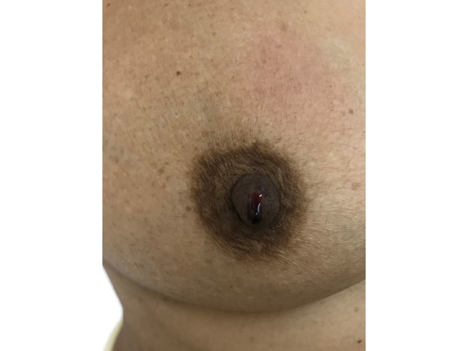
| Clinical presentation: | Woman aged 70 years with left breast carcinoma who underwent left MRM in 1994. She presented in 2015 with right breast serous nipple discharge. The nipple discharge is occasional, serous for 1 year, with one episode of blood-stained discharge 1 day ago. |
 |
Mammography:
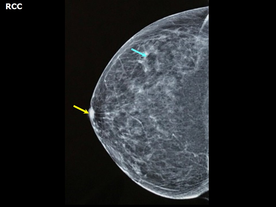 | 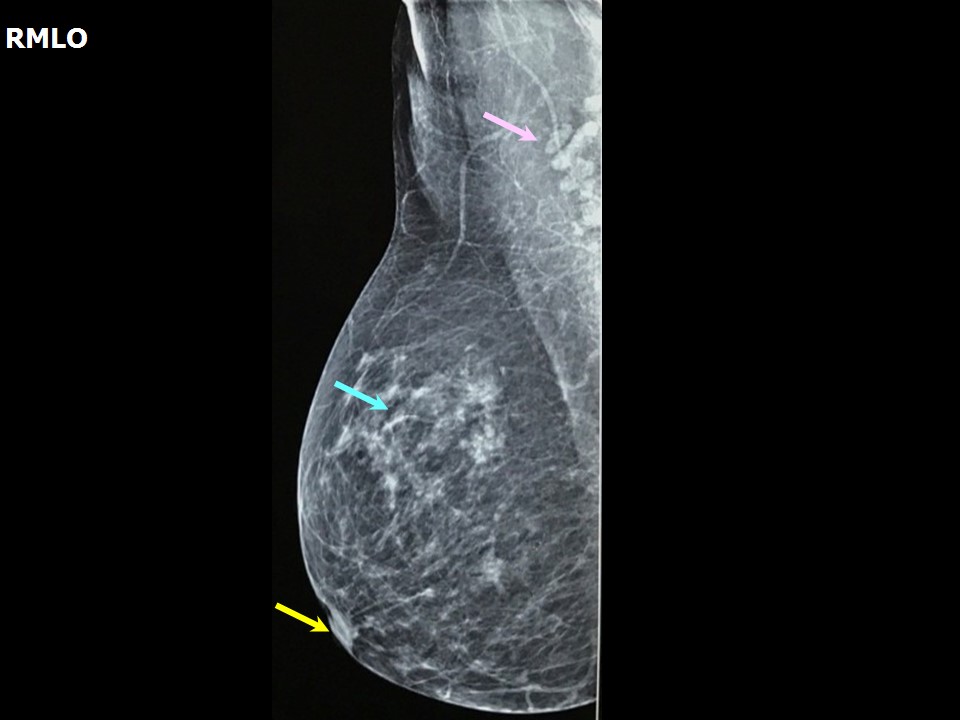 |
| Breast composition: | ACR category b (there are scattered areas of fibroglandular density) | Mammography features: |
| ‣ Location of the lesion: | Right breast |
| ‣ Mass: | |
| • Number: | 0 |
| • Size: | None |
| • Shape: | None |
| • Margins: | None |
| • Density: | None |
| ‣ Calcifications: | |
| • Typically benign: | Round, vascular |
| • Suspicious: | None |
| • Distribution: | None |
| ‣ Architectural distortion: | None |
| ‣ Asymmetry: | None |
| ‣ Intramammary node: | None |
| ‣ Skin lesion: | None |
| ‣ Solitary dilated duct: | None |
| ‣ Associated features: | None |
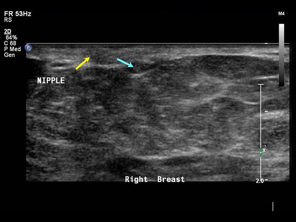
Ultrasound:
 | 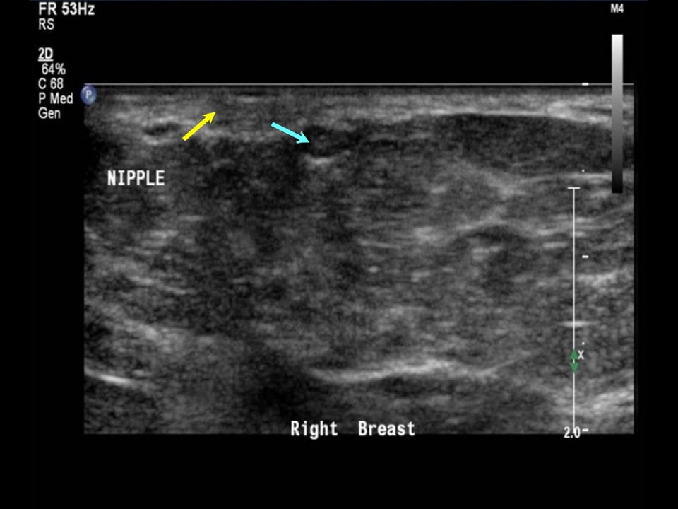 |
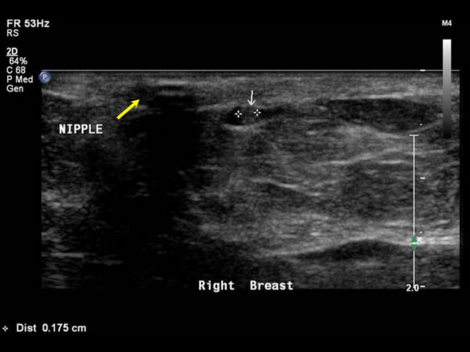 |
| Ultrasound features: Right breast, lower inner quadrant at 4 oclock | |
| ‣ Mass | |
| • Location: | Right breast, lower inner quadrant at 4 oclock |
| • Number: | 1 |
| • Size: | 0.1 cm within dilated duct |
| • Shape: | Round |
| • Orientation: | Parallel |
| • Margins: | Circumscribed |
| • Echo pattern: | Isoechoic |
| • Posterior features: | No posterior features |
| ‣ Calcifications: | None |
| ‣ Associated features: | Duct changes |
| ‣ Special cases: | Solitary dilated duct |
BI-RADS:
BI-RADS Category: 4B (moderate suspicion of malignancy)Further assessment:
Further assessment advised: Further imaging with breast MRIMRI:
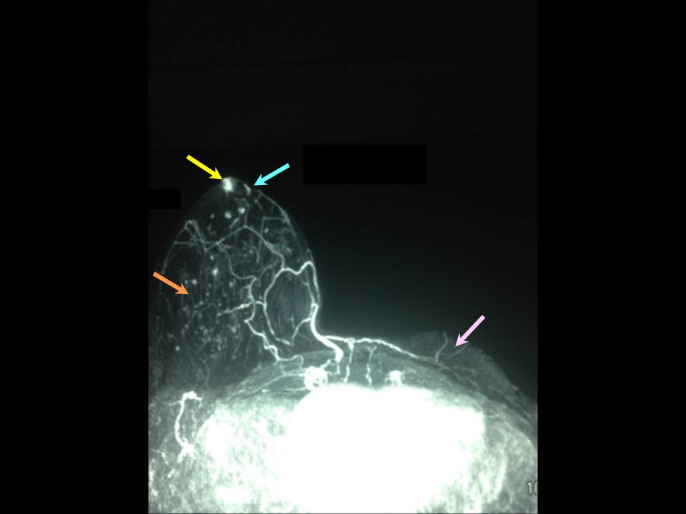 | 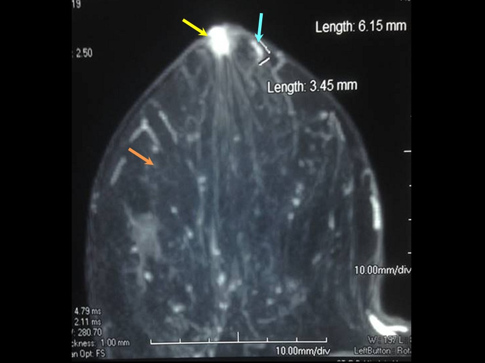 |
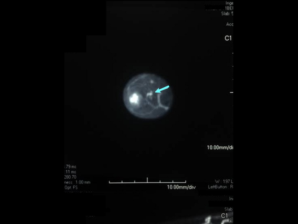 | 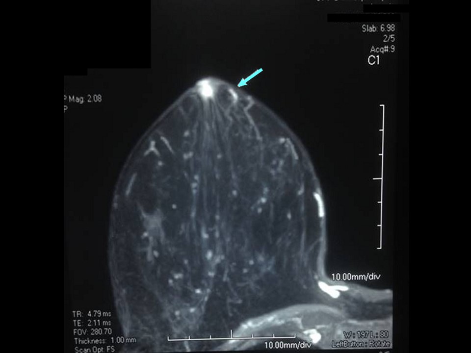 |
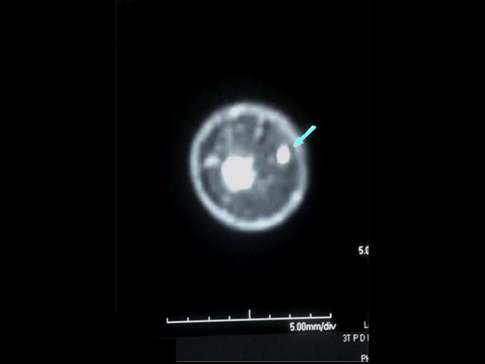 |
| MRI features: | ||
| ‣ MRI features: | Amount of fibroglandular tissue: ACR category a (the breasts are almost entirely fatty). Background parenchymal enhancement: Minimal (< 25%), symmetrical | |
| ‣ Location: | Right breast at 3 oclock | |
| ‣ Focus: | No | |
| ‣ Mass: | ||
| • Shape: | Tiny linear | |
| • Margin: | Circumscribed | |
| • Internal enhancement: | Homogeneous | |
| • Kinetic curve: | Type 3 | |
| ‣ Non-mass enhancement: | ||
| • Distribution: | Ductal | |
| • Internal enhancement: | Homogeneous | |
| ‣ Non-enhancing findings: | No | |
| ‣ Associated features: | No | |
| ‣ Axillary nodes: | No | |
Case summary:
| Postmenopausal woman presented with right breast blood-stained nipple discharge, diagnosed as focal short-segment solitary prominent duct with intraductal lesion, BI-RADS category 4B on imaging and as papilloma on histopathology. |
Learning points:
|